CG vs 2D Echo vs Stress Test: Which Heart Test Do You Need
Table of Contents
TogglePawana Hospital Team
By Pawana Hospital Team | Gynecology & Endocrinology | August 2026 | 9 min read
Heart-related symptoms can be confusing. Chest discomfort, shortness of breath, palpitations, unusual tiredness, or dizziness may have several possible causes, and symptoms alone cannot always tell doctors exactly what is happening inside the heart.
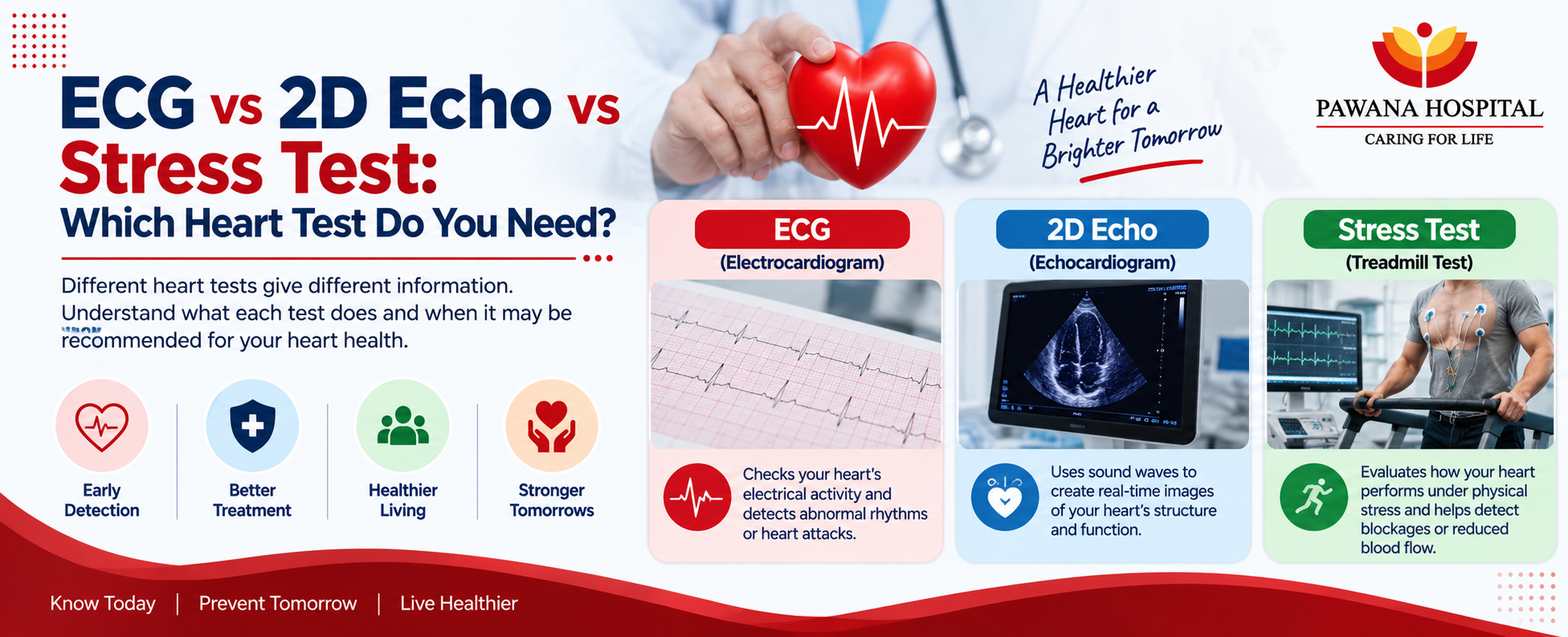
This is why cardiologists use different diagnostic tests to evaluate different aspects of heart health. Three commonly recommended cardiac tests are the ECG (Electrocardiogram), 2D Echo (2D Echocardiogram), and Stress Test or TMT (Treadmill Test).
Although all three examine the heart, they do very different jobs. An ECG primarily examines the heart’s electrical activity and rhythm.
A 2D Echo uses ultrasound to assess the heart’s structure and pumping function. A stress test evaluates how the heart responds when its workload increases during exercise. Understanding these differences can help patients know why a particular test has been recommended and why, in some cases, more than one cardiac investigation may be necessary.
Introduction – Why Different Heart Tests Are Needed
The heart is both a muscular pump and an electrical organ. It has chambers and valves, an electrical conduction system that controls the heartbeat, and coronary arteries that supply blood to the heart muscle.
Because heart disease can affect any of these systems, there is no single test that can identify every cardiac condition. For example, a patient may have an abnormal heart rhythm that is visible on an ECG but no major structural abnormality.
Another person may have a valve or heart-muscle problem that is better evaluated with a 2D Echo. Some heart problems may become more noticeable only when the heart is working harder during exercise, which is where a stress test may provide useful information.
A cardiologist chooses investigations based on the patient’s symptoms, age, medical history, examination findings, cardiovascular risk factors, and results of previous tests.
What Is an ECG?
An Electrocardiogram (ECG or EKG) is a simple test that records the electrical activity generated by the heart.
Every heartbeat is controlled by electrical signals. An ECG records these signals through electrodes attached to the skin and displays them as wave patterns that a doctor can interpret.
The test can provide information about:
- Heart rate Heart rhythm
- Electrical conduction through the heart
- Certain abnormal rhythms or arrhythmias
- Possible evidence of current or previous heart-muscle injury
- Certain changes that may suggest reduced blood supply to the heart
An ECG is quick and non-invasive. It is commonly one of the first investigations performed when someone presents with chest discomfort, palpitations, dizziness, breathlessness, or suspected heart-related symptoms.
However, an ECG records the heart only during the short period when the test is being performed. Some intermittent abnormalities may therefore not appear on a routine ECG.
What Is a 2D Echo?
A 2D Echocardiogram, commonly called a 2D Echo, is an ultrasound examination of the heart.
Instead of measuring electrical activity, it uses sound waves to create moving images of the heart. These images allow the cardiologist to examine how the heart is structured and how effectively it is functioning.
A 2D Echo can help doctors assess:
- Heart chambers
- Heart valves
- Heart-wall movement
- Pumping ability of the heart
- Heart-muscle function
- Certain congenital structural abnormalities
- Fluid around the heart
- Some changes associated with previous cardiac damage
One important measurement obtained during an echocardiogram is the ejection fraction (EF), which helps assess how effectively the heart’s main pumping chamber is pumping blood.
Because an ECG and 2D Echo examine different aspects of the heart, a normal ECG does not automatically mean that a 2D Echo will also be normal.
What Is a Stress Test (TMT)?
A Stress Test, commonly called a Treadmill Test (TMT), evaluates how the heart performs when its workload increases.
During the test, the patient usually walks on a treadmill while healthcare professionals monitor the ECG, heart rate, blood pressure, symptoms, and response to progressively increasing exercise.
A stress test may be considered when doctors want to assess:
• Exercise-related chest discomfort
•Exercise tolerance
•ECG response during exertion
•Symptoms that appear during physical activity
•Possible evidence of reduced blood flow to the heart during exercise
•Functional capacity in selected cardiac patients
A TMT is not suitable for every patient. A cardiologist should determine whether it is appropriate based on symptoms, medical history, resting ECG findings, physical ability, and overall cardiovascular risk.
ECG vs 2D Echo vs Stress Test – Key Differences
Although these tests are often discussed together, they evaluate different aspects of cardiac health.
ECG Main purpose:
Evaluates the electrical activity of the heart. It provides information about heart rhythm, rate, electrical conduction, and certain changes associated with heart-muscle stress or damage.
2D Echo Main purpose:
Evaluates the heart’s structure and function. It allows doctors to see the chambers, valves, heart walls, and pumping action of the heart.
Stress Test / TMT Main purpose:
Evaluates the heart’s response to physical exertion. It helps doctors observe how the ECG, heart rate, blood pressure, and symptoms change when the heart has to work harder.
Therefore, these tests should not be considered interchangeable. The most appropriate test depends on what the doctor is trying to investigate.
What Can Each Heart Test Detect?
Understanding what each investigation can reveal makes the differences easier to understand.
ECG May Help Identify:
• Abnormal heart rhythms Fast or slow heart rate
• Certain electrical conduction abnormalities
• Changes that may occur during a heart attack
• Evidence suggesting previous heart-muscle injury
• Certain changes associated with reduced blood supply
2D Echo May Help Identify:
• Heart valve abnormalities
• Reduced heart pumping function
• Abnormal heart-wall movement
• Enlargement of heart chambers
• Certain structural heart conditions
• Fluid around the heart Some congenital heart abnormalities
Stress Test May Help Evaluate:
• Symptoms occurring during exercise
• Exercise tolerance
• Heart-rate response to physical activity
• Blood-pressure response during exercise
• ECG changes occurring under physical stress
• Possible exercise-related reduction in blood flow to the heart
No test should be interpreted in isolation. Results should be evaluated by a doctor together with symptoms, risk factors, examination findings, and other investigations.
Which Test Is Recommended for Chest Pain?
Chest pain is one of the most common reasons people undergo cardiac testing, but there is no universal test that is appropriate for every case.
When a person develops new, severe, persistent, or suspicious chest pain, an ECG may be one of the first cardiac investigations performed, particularly when doctors need to evaluate a possible acute heart problem.
Depending on the situation, additional investigations such as blood tests, echocardiography, coronary imaging, angiography, or other tests may also be required.
For stable symptoms that occur primarily during physical exertion, a cardiologist may consider a stress test when it is medically appropriate.
A 2D Echo may be recommended when the doctor wants to assess the heart’s pumping function, valves, wall movement, or other structural features.
Most importantly, someone experiencing severe chest pressure, pain spreading to the arm or jaw, sudden breathlessness, cold sweating, fainting, or other possible heart-attack symptoms should seek emergency medical care rather than waiting to arrange a routine heart test.
Which Test Is Used for Palpitations or Breathlessness?
The appropriate investigation depends on what is causing the symptom.
For Palpitations
An ECG is commonly used as an initial investigation because palpitations may be associated with abnormal heart rhythms.
However, if the palpitations occur intermittently and are not present during the ECG, the doctor may recommend longer-duration heart-rhythm monitoring.
For Breathlessness
A 2D Echo may be useful when doctors need to evaluate heart pumping function, valves, chamber size, or other structural causes that could contribute to breathlessness.
Depending on whether symptoms occur during physical activity, a stress test may sometimes be considered. Breathlessness can also result from conditions unrelated to the heart, including respiratory disease, anaemia, and other medical problems.
A clinical evaluation is therefore important before deciding which test is required.
Do You Need More Than One Heart Test?
Yes, sometimes.
An ECG, 2D Echo, and stress test provide different types of information, so one investigation may not answer every clinical question.
For example, a patient may have an ECG to evaluate heart rhythm and electrical activity, followed by a 2D Echo to examine heart structure and pumping function.
If symptoms mainly occur during exercise, the cardiologist may subsequently consider a stress test. Similarly, abnormal findings on one test may lead to additional investigations for a more detailed assessment.
The goal is not to perform every heart test on every patient.
The goal is to select investigations that provide the information needed for that individual’s symptoms and cardiovascular risk.
What Happens During These Tests?
Understanding what to expect can make cardiac testing less stressful.
During an ECG
Small adhesive electrodes are placed on the chest and usually on the limbs.
These electrodes connect to the ECG machine and record the heart’s electrical signals. The recording itself generally takes only a short time and does not involve electrical current being sent into the body.
During a 2D Echo
The patient usually lies on an examination bed while a healthcare professional moves an ultrasound probe over different areas of the chest.
A special gel is applied to help obtain clear ultrasound images. The images allow the doctor to observe the heart while it is beating.
During a Stress Test
Electrodes are attached to the chest, and blood pressure and ECG are monitored while the patient walks on a treadmill.
The intensity is gradually increased according to the testing protocol while healthcare professionals observe symptoms and cardiovascular responses.
The test may be stopped if the target level is reached, the patient develops concerning symptoms, or the medical team identifies a reason to stop.
Who Should Consider a Cardiac Evaluation?
You do not necessarily need to wait for severe symptoms before discussing your heart health with a doctor.
A cardiac evaluation may be appropriate if you:
• Experience recurring chest discomfort
•Have unexplained shortness of breath
• Experience frequent palpitations
•Feel dizzy or faint without a clear reason
• Notice a significant decline in exercise tolerance
• Have high blood pressure Have diabetes Have high cholesterol
•Smoke or have smoked previously
• Have a strong family history of heart disease
•Have previously abnormal cardiac test results
People with multiple cardiovascular risk factors may benefit from discussing preventive heart evaluation with their doctor even when they do not have obvious symptoms
Cardiology & Heart Testing at Pawana Hospital
Heart symptoms can range from minor concerns to medical emergencies, which makes accurate and timely evaluation important.
At Pawana Hospital, cardiac patients can receive evaluation through the hospital’s Cardiology Department.
Depending on the patient’s symptoms and clinical condition, cardiac assessment may include ECG, 2D Echo, laboratory investigations, and other diagnostic tests recommended by the cardiologist.
For patients who require more advanced evaluation or treatment, Pawana Hospital also provides cardiac services including coronary angiography and angioplasty through its cardiac care facilities.
The appropriate investigation is selected after considering the patient’s symptoms, medical history, cardiovascular risk factors, examination findings, and previous reports.
If you experience persistent or recurring cardiac symptoms, consulting a cardiologist can help determine which investigation is appropriate rather than choosing a heart test on your own.
Frequently Asked Questions (FAQs)
1. Which is better: ECG or 2D Echo?
Neither test is universally better because they evaluate different aspects of the heart. An ECG primarily assesses electrical activity and rhythm, whereas a 2D Echo evaluates heart structure, valves, wall movement, and pumping function.
2. If my ECG is normal, do I still need a 2D Echo?
Possibly. A normal ECG does not rule out every structural or functional heart condition. A cardiologist may recommend a 2D Echo depending on your symptoms, examination findings, and medical history.
3. Can an ECG detect every heart blockage?
No. An ECG can provide important information and may show changes associated with reduced blood flow or a heart attack, but a normal ECG does not exclude all coronary artery disease. Additional testing may be required when clinically indicated.
4. Can a 2D Echo detect blocked coronary arteries?
A standard 2D Echo primarily evaluates the structure and function of the heart rather than directly visualising coronary artery blockages. It may reveal changes in heart-wall movement or pumping function that provide additional clinical information, but other investigations may be needed to evaluate the coronary arteries.
5. Is a TMT the same as an ECG?
No. Although ECG monitoring is used during a treadmill test, a resting ECG records the heart while you are at rest. A TMT evaluates cardiovascular responses while the workload gradually increases during exercise.
6. Is a stress test safe?
Stress testing is generally performed under medical supervision with continuous monitoring. However, it is not appropriate for everyone. A doctor should first determine whether the test is suitable based on your health and cardiac condition.
Conclusion:
ECG, 2D Echo, and stress testing are valuable cardiac investigations, but they answer different questions.
An ECG examines the electrical activity and rhythm of the heart. A 2D Echo examines its structure and pumping function.
A Stress Test evaluates how the heart responds when its workload increases during exercise. The right test therefore depends on your symptoms, medical history, risk factors, and what your cardiologist needs to evaluate.
If you experience recurring chest discomfort, unexplained breathlessness, palpitations, dizziness, or reduced exercise capacity, do not simply choose a test based on symptoms found online.
A cardiac consultation can help determine which investigation — or combination of investigations — is most appropriate.
And if chest pain is severe, persistent, or accompanied by breathlessness, sweating, fainting, or pain spreading to the arm, back, neck, or jaw, seek emergency medical attention immediately.