Pneumonia in Adults: Comprehensive Guide to Symptoms, Pathophysiology, Diagnosis & Management
Table of Contents
Toggle1. Introduction & Global Health Burden
Pneumonia remains one of the leading causes of lower respiratory tract infection morbidity and mortality worldwide among adults. Characterized by an acute inflammatory exudative response within the pulmonary parenchyma, it affects millions of patients annually and places a substantial burden on healthcare systems.
Pneumonia spans a broad clinical spectrum ranging from mild out-patient infection (“walking pneumonia”) to rapidly progressive, life-threatening respiratory failure requiring intensive care unit (ICU) admission and mechanical ventilation.
2. Anatomic Pathophysiology
The lower respiratory tract is normally maintained in a sterile state through anatomical barriers, mucociliary clearance, cough reflexes, and alveolar macrophage phagocytosis.
Pneumonia develops when pathogenic micro-organisms bypass these defense mechanisms:
Alveolar Infiltration:
Neutrophils and serum proteins cross the pulmonary capillary membrane into the alveoli, leading to dense consolidation.
Impaired Gas Exchange:
Fluid and cellular debris fill the air sacs, causing intra-pulmonary ventilationperfusion (V/Q) mismatch and arterial hypoxemia.
Consolidation:
Infection spreads to adjacent alveoli, potentially consolidating an entire lobe (Lobar Pneumonia) or presenting as patchy infiltrates (Bronchopneumonia).
3. Clinical Classifications
| Category | Acronym | Setting & Pathogen Profile |
|---|---|---|
| Community-Acquired Pneumonia | CAP | Contracted outside healthcare facilities; predominantly caused by Streptococcus pneumoniae or respiratory viruses. |
| Hospital-Acquired Pneumonia | HAP | Develops ≥48 hours after admission; frequently involves resistant pathogens like Pseudomonas aeruginosa or MRSA. |
| Ventilator-Associated Pneumonia | VAP | Arises >48 hours post-intubation; associated with micro-aspiration in ICU settings. |
| Aspiration Pneumonia | AP | Occurs following inhalation of foreign contents; common in patients with dysphagia or altered consciousness. |
4. Etiological Pathogens & High-Risk Factors
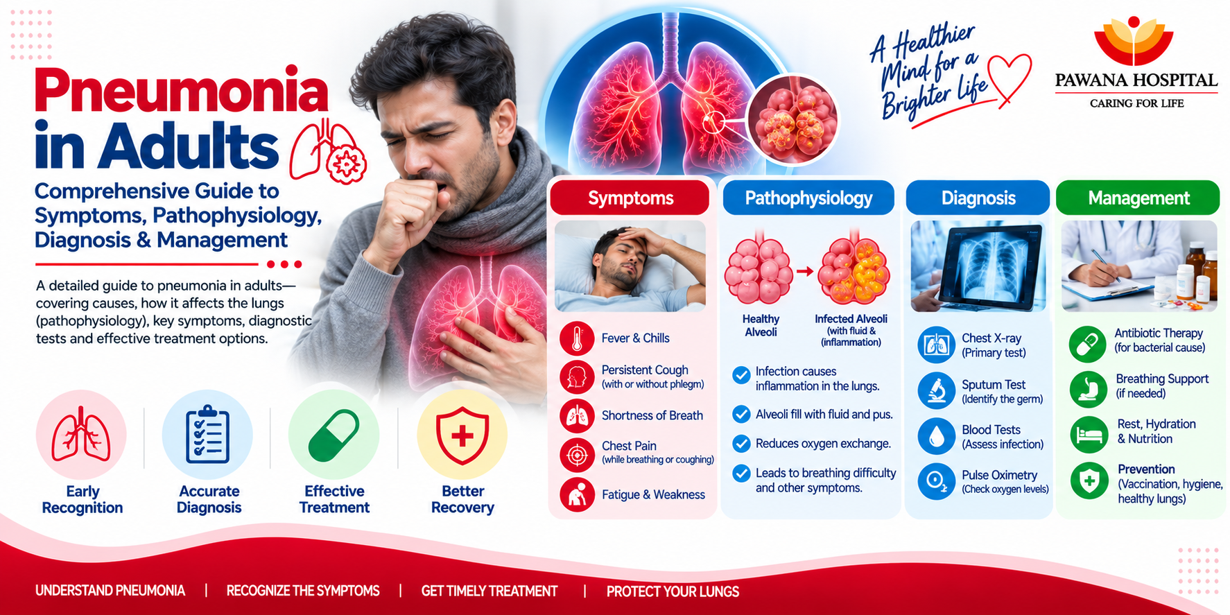
Infectious agents causing pneumonia include bacterial pathogens (Streptococcus pneumoniae, Haemophilus influenzae), atypical organisms (Mycoplasma pneumoniae, Legionella), and viral agents (Influenza, RSV, SARS-CoV-2).
| Risk Category | Clinical Risk Factors | Underlying Mechanism |
|---|---|---|
| Age & Senescence | Adults aged ≥ 65 years | Immunosenescence and diminished cough reflex. |
| Chronic Comorbidities | COPD, Asthma, Heart Failure, Diabetes | Impaired mucociliary clearance and immune dysfunction. |
| Immunosuppression | Chemotherapy, long-term steroids | Reduced cellular pulmonary immune surveillance. |
| Lifestyle Exposures | Active smoking, heavy alcohol use | Destruction of ciliated cells and macrophage dysfunction. |
5. Clinical Symptoms & Diagnostic Protocol
Symptoms range from acute high-grade fever, chills, cough with purulent sputum, and pleuritic chest pain to atypical presentations. Elderly patients often present with confusion, lethargy, or hypothermia without fever.
| Diagnostic Modality | Target Objective & Clinical Significance |
|---|---|
| Chest Radiograph (X-Ray) | Confirms lobar consolidation, interstitial infiltrates, or pleural effusion. |
| High-Resolution CT (HRCT) | Evaluates complex non-resolving cases, cavitation, or empyema. |
| Pulse Oximetry & ABG | Quantifies hypoxemia, hypercapnia, and arterial acid-base balance. |
| Biomarkers & Cultures | Serum Procalcitonin (>0.25 ng/mL) and Sputum/Blood cultures guide therapy. |
6. CURB-65 Clinical Risk Stratification Score
The CURB-65 score determines whether a patient requires outpatient care, ward hospitalization, or intensive care unit (ICU) admission.
| Parameter | Clinical Threshold | Points |
|---|---|---|
| C — Confusion | New disorientation in time, place, or person | 1 Point |
| U — Urea | Blood Urea Nitrogen (BUN) > 19 mg/dL (>7 mmol/L) | 1 Point |
| R — Respiratory Rate | ≥30 breaths per minute | 1 Point |
| B — Blood Pressure | Systolic BP < 90 mmHg OR Diastolic BP ≤ 60 mmHg | 1 Point |
| 65 — Age | Age ≥ 65 years | 1 Point |
Score Triage: 0–1 = Low Risk (Outpatient); 2 = Moderate Risk (Inpatient Ward); 3–5 = Severe Risk (Evaluate for ICU Admission).
7. Evidence-Based Therapeutic Management Outpatient CAP Therapy:
Amoxicillin 1g TID OR Macrolide (Azithromycin) OR Doxycycline 100mg BID. Comorbid patients receive Beta-lactam plus Macrolide or Respiratory Fluoroquinolone.
Inpatient Hospital Therapy:
IV Beta-lactam (Ceftriaxone 1-2g daily) PLUS IV Macrolide or Fluoroquinolone.
ICU / HAP Therapy:
Broad-spectrum anti-pseudomonal coverage (Piperacillin-Tazobactam or Meropenem) plus MRSA coverage (Vancomycin) adjusted upon culture reports.
Supportive Oxygen Therapy:
Maintained via high-flow nasal cannula or Venturi mask to achieve SpO2 of 92%–96% (88%–92% in COPD).
8. Recovery Timeline
| Time Frame | Clinical Expectation |
|---|---|
| 1 – 3 Days | Fever defervescence and vital sign stabilization. |
| 1 – 2 Weeks | Substantial decrease in cough, chest pain, and sputum output. |
| 6 – 12 Weeks | Resolution of fatigue and complete radiographic clearance on X-ray. |
9. Prevention & Immunization Protocols
Preventive strategies reduce the incidence and mortality of adult pneumonia significantly:
Pneumococcal Vaccination:
Sequential PCV15 / PCV20 and PPSV23 administration for adults ≥65 years and younger individuals with high-risk conditions.
Annual Influenza Immunization:
Reduces secondary bacterial post-viral pneumonia risks by up to 60%.
Smoking Cessation & Hygiene:
Protects mucociliary clearance and alveolar macrophage health.
10. Frequently Asked Questions (FAQs)
1. What is the difference between “walking pneumonia” and severe pneumonia?
“Walking pneumonia” is a mild atypical pneumonia (often Mycoplasma) where hospital care is not required. Severe pneumonia causes major systemic distress requiring medical admission.
2. Why is a follow-up Chest X-ray necessary after recovery?
A repeat X-ray is performed 6 to 8 weeks post-recovery in smokers or adults over 50 to confirm complete resolution and rule out underlying lesions.
3. Are antibiotics effective against viral pneumonia?
No, antibiotics target bacteria. However, antivirals may be used, and antibiotics added if a secondary bacterial infection develops.
11. Summary & Conclusion
Pneumonia is a serious respiratory infection requiring timely clinical recognition, diagnostic precision, and targeted management. Through clinical evaluation, severity scoring, and robust vaccination programs, patients can achieve effective recovery. Your lung health deserves professional care. Consult the Department of Pulmonology at Pawana Hospital for specialized medical guidance.